
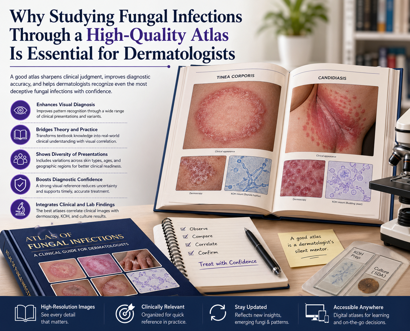
Why Studying Fungal Infections Through a High-Quality Atlas Is Essential for Dermatologists
The Challenge of Diagnosing Fungal Skin Infections
Fungal infections are among the most common conditions encountered in dermatology, yet they remain surprisingly difficult to diagnose with precision. Many fungal skin infections closely resemble other dermatologic conditions such as eczema, psoriasis, or bacterial infections. This overlap makes visual diagnostic skills essential for every dermatologist.
To improve accuracy and confidence, dermatologists increasingly rely on a high-quality dermatology atlas—a critical tool for mastering fungal infection diagnosis.
The Role of a Dermatology Atlas in Clinical Practice
A dermatology atlas is far more than a collection of images. It is a visual diagnostic guide designed to enhance pattern recognition, which lies at the heart of dermatology.
Subtle variations in:
- Skin color
- Scaling patterns
- Lesion borders
- Distribution across the body
can completely change a diagnosis. A high-quality atlas provides comprehensive image collections, including both classic presentations and atypical variants of fungal infections. This helps clinicians sharpen their observational skills beyond theoretical learning.
Bridging the Gap Between Theory and Real-World Diagnosis
Medical textbooks often describe conditions like tinea corporis, tinea versicolor, or cutaneous candidiasis in technical terms. However, without strong visual correlation, these descriptions can remain abstract.
A dermatology atlas bridges this gap by:
- Translating theory into real clinical images
- Helping dermatologists quickly recognize fungal infection patterns
- Improving diagnostic speed in clinical settings
This is especially valuable in busy clinics where rapid and accurate decisions are essential.
Understanding Variability in Fungal Infections
Fungal infections do not present the same way in every patient. Their appearance can vary depending on:
- Skin type and pigmentation
- Immune status
- Geographic region
- Environmental and cultural factors
A comprehensive atlas exposes dermatologists to this diversity, reducing the risk of misdiagnosis—particularly in regions where fungal infections are highly prevalent.
Improving Diagnostic Confidence and Patient Outcomes
Consistent use of a high-quality atlas helps dermatologists build a mental library of clinical images. This leads to:
- Faster recognition of fungal infections
- Increased diagnostic confidence
- More accurate treatment decisions
Ultimately, this improves patient outcomes, reduces unnecessary treatments, and minimizes diagnostic errors.
Key Features of a High-Quality Dermatology Atlas
Not all atlases provide the same value. The best dermatology atlases for fungal infections should include:
- High-resolution clinical images
- Clear annotations and descriptions
- Dermoscopic imaging when applicable
- Correlation with laboratory findings (e.g., KOH examination, fungal culture)
- Regular updates reflecting emerging fungal species and resistance patterns
Choosing the right atlas is essential for staying current in modern dermatology practice.
Digital Atlases and Modern Learning
With the rise of digital tools, many dermatology atlases are now available as:
- Mobile applications
- Online databases
- Interactive learning platforms
These resources allow dermatologists to access visual references instantly during patient consultations, making them an indispensable part of modern clinical workflows.
Developing the Dermatologist’s Eye
Mastering fungal infections in dermatology is not just about memorizing disease names or treatments—it is about developing a highly trained eye for visual diagnosis.
A high-quality dermatology atlas acts as a silent mentor, guiding clinicians through complex and often deceptive presentations of fungal infections. With consistent use, it becomes one of the most powerful tools for improving diagnostic accuracy and delivering better patient care.